1. DIAGNOSTIC SERVICES:
• VISUAL ACUITY CHARTS : To quantify vision
• AUTO REFRACTOMETER AND KERATOMETER: To calculate amount of
refractive errors and corneal curvature.
• NCT & SHIOTZ TONOMETER: To measure intra ocular pressure.
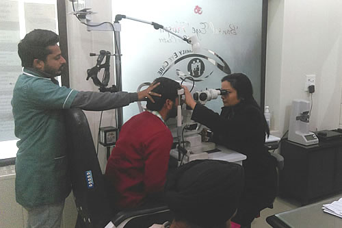
• SLIT LAMPS: To diagnose Anterior and Posterior abnormalities
of eye.
• DIRECT AND INDIRECT OPHTHALMOSCOPES: To diagnose retinal
diseases.
• DIGITAL FUNDUS CAMERA: For Fluorecsein Angiography to diagnose
retinal disorders.
• GONIOSCOPE: To assess anterior chamber angles in glaucoma or
trauma patients.
• PERIMETRY: To assess visual fields in Glaucoma and Neuro-Ophthalmology
patients.
• BIOMETRY: To calculate IOL power.
• PACHYMETRY: To assess corneal thickness for treating Glaucoma
and LASIK patients.
• SYNOPTOPHORE: For evaluation and treatment of squint patients.
2. LASER TREATMENT SERVICES:
• NdYAG LASER : For Cataract and Glaucoma patients.
• GREEN LASER: For treatment of retinal disorders.
• LASIK LASER: For treatment of refractive errors.
3. MINOR TREATMENT SERVICES:
• Dressings.
• Syringing and probing of lacrimal sac.
• Stitch removal.
• Subconjunctival / retrobulbar/ peribulbar injections.
• Intra lesional steroid injections(chalazion).
• Sub tenon injections.
• Intra vitreal injections.
4. SURGICAL SERVICES:
Various surgeries & LASER Procedures being performed in
operation theatre of the hospital are as under:
CATARACT SERVICES
1. Clouding of Natural crystalline clear Lens Inside the eye is
termed as cataract.
2. It is an age related change and usually seen in people above
50 years of age.
3. Less common causes include injury, metabolic diseases,
inflammation in the eye etc.
4. Cataract blurs the vision since it causes transmission block
for light rays.
5. It leads to loss of quality vision.
6. Cataract can develop faster in one or both the eyes.
7. Once the cataract is removed there is no scope for the
cataract to develop again.
8. Once the cataract is detected surgery is the only Ideal
treatment.
9. Sometimes the blurring of vision occurs due to thickening of
the supporting membrane of the IOL (posterior capsular opacity)
it can be corrected with YAG Laser Capsulotomy.
Phacomulsification is the Latest technique to remove the
cataract. Advantages:
1. No Hospitalization
2. No Injection
3. No stitches
4. Early visual recovery and rehabilitation
5. Totally painless
6. Done through a tiny self sealing tunnel incision
7. Less Post – operative precautions
IOL (Intra Ocular Lens)
1. After cataract removal eye loses its focusing power. In order
to restore focusing power an intra-ocular lens has to be
implanted. If not, thick high powered glasses are required.
2. Advanced IOL’s like foldable Uni-focal, Multifocal, Aspheric,
Aberration free and Toric IOL’s will be implanted for quality
vision.
a) In Uni-focal IOL implantation, reading glasses will be
prescribed.
b) In Multi-focal IOL implantation both for distance and near,
dependency on the glasses will be reduced.
GLAUCOMA SERVICES
Glaucoma is described as progressive optic atrophy leading to
gradual loss of vision due to multiple factors. Intra Ocular
pressure (IOP) is one of the factors that affect the functioning
of optic nerve.
Mainly Glaucoma is divided into two groups
1. Congenital Glaucoma:- This is seen in newborn infants. It is
due to developmental defect of draining channels and the front
portion of the eye ball. It is a serious sight threatening
problem. If diagnosed in early stage can be treated by surgery
&/or medications.
2. Acquired Glaucoma: Again it can be classified as:
a) Primary Glaucoma:- Which occurs without any underlying cause.
It is due to the age and changes in anatomical configuration. It
can again be classified as:-
i) Open angle Glaucoma: This usually occurs in people above 40
Years. Though the drainage area is open, the draining channels
are blocked due to age related changes.
ii) Narrow angle Glaucoma:- The irido-corneal angle is narrow
which causes obstruction to the outflow of fluid in the eye
ball. This increases the intraocular pressure which in turn
hampers the optic nerve functioning.
b) Secondary Glaucoma:- is due to the underlying causes like
injury, inflammation, diabetes etc.
People at Risk- Those:
1. Having refractive errors specially High Myopia
2. With family history of Glaucoma.
3. With history of eye injuries.
4. Using steroid eye drops.
5. Diabetics and hypertensives.
Points to Remember
1. Glaucoma is a sneak thief of sight since no symptoms will be
observed.
2. Regular screening after 40 years of age is a must.
3. It runs in the family.
4. Glaucoma cannot be cured but can be maintained with regular
follow-ups and medication.
5. Glaucomatous optic nerve damage is irreversible.
6. Glaucoma treatment is life time commitment.
7. Diagnosis is based on the reports of Intraocular Pressure
(IOP) measurement, visual field test, Optic nerve head
examination and Gonioscopy.
8. Early diagnosis and treatment will save the vision.
Our Glaucoma clinic is equipped with
1. Tono meters- Applanation , Schiotz, NCT, Tono pen : to
measure IOP
2. Slit lamp examination and Gonioscopy: To assess the drainage
area of the eye (angle of the anterior chamber)
3. Automated perimeter : to evaluate the visual field defect
which indicates the extent of optic nerve damage.
4. OCT: It is a scanning device where the structural integrity
of optic nerve and retina is evaluated in relation to Glaucoma.
Treatment
Treatment depends on the type and stage of Glaucoma. Most of the
time it is the medical line of treatment. Eye drops will be used
to lower and maintain IOP and as neuroprotectors. Other
treatments include:
1. Surgical: Filtering Surgery (Trabeculectomy)
2. YAG LASER procedure: To perform YAG LASER Iridotomy in narrow
angle Glaucoma.
3. Argon LASER trabeculoplasty and Gonio photocoagulation as a
palliative treatment in Glaucoma.
4. Cylcocryopexy - Cylcophotocoagulation: is a destructive
procedure to freeze the ciliary body and to reduce the aqueous
humor production thereby reducing the IOP in painful blind eyes.
“We offer comprehensive Glaucoma eye checkup at Bansal Eye
Hospital with state of art diagnostic and treatment modalities.”
OCULOPLASTY
It is the reconstruction of the eye ball, lids and orbit without
disturbing cosmoses under this, the following treatments will be
given:
Squint
1. Squint is the condition where, a deviation of the eye from
its original axis while fixing the focus on given object, is
observed
2. It can affect one or both the eyes.
3. It is very important to observe this condition in the new
born infants, preschool children and school going children.
4. Though Squint is a disorder of ocular muscle balance, it can
also occur by various other reasons e.g. low vision in one eye,
trauma – etc.
5. If squint is not treated in time before the age of 8 Years,
in a child especially when there is unilateral squint, blunting
of vision occurs permanently. This is called as Amblyopia.
6. At Bansal Eye Hospital we periodically organize eye check-ups
for school children to pick up this problem at an early stage
and treat them with spectacle corrections, Pleoptic-exercises,
anti suppression treatment (patching therapy) and/or surgery.
7. Squint can also occur in adults due to trauma, neurological
disorders and accordingly treatment is given.
Blepharoplasty
• Various eyelid disorders like Ptosis (Dropping of eye lid)
Entropion, Ectropion, cosmetic lid surgery following eye lid
trauma are performed regularly.
• Tarsorraphy and gold implant surgery will be done for
logophthamos following facial nerve palsy.
Botox Injection
Botulinum injections are used in essential blepharospasm to
relax the eyelid muscles, to reduce diplopia, to reduce severity
of corneal dryness following lagophtholmos and for cosmetic
reasons.
DCR
Constant watering in one or both eyes related to obstruction in
the tear passage can be corrected by DCR:- A procedure which
will create opening from tears sack to nasal cavity. It can be
done by either manual method or laser method.
OTHERS
We at Bansal Eye Hospital offer the above treatment with our
team of experienced and well trained doctors in the field .
• Oculoplasty department is well equipped with radio frequency
cautery, Micro surgical instruments implants.
• Amniotic Membrane Grafting for recurrent Pterygium/ corneal
Ulcers etc. Tissue fibrin GLUES e.g. BAXTER for various
pathologies.
• Orbital Blow out Fractures – Repair & floor Reconstruction
with Medpore implants.
VITREO- RETINA SERVICES
Diabetic Retinopathy
Diabetic Retinopathy is a disorder of the retina which affects
most of the people with Diabetes Mellitus leading to visual
disturbance, at times total blindness. Diabetic Mellitus is a
condition where the blood sugar levels are above the normal
range due to absence or deficiency of insulin in our body.
There are two types of Diabetes
1. IDDM (Insulin Dependent Diabetic Mellitus)
2. NIDDM (Non-insulin Dependent Diabetic Mellitus)
• Diabetic Retinopathy is known to be more common in IDDM. It
affects the small capillaries of the retina thereby damaging the
nerve cells leading to vision problem.
• The wall of the capillaries become weak and starts leaking
plasma fluid and fat leading to retinal oedema.
• Ballooning of capillaries i.e. Aneurysms is a common feature.
• The part of retina deprived of blood supply starts developing
new abnormal vessels which break frequently causing retinal and
pre-retinal haemorrhages. If this happens in the central part of
retina- it is known as Maculopathy, which disturbs the vision to
a greater extent.
• Usually, Diabetes Mellitus does not affect the eye in the
initial period (approx. 5-6 years) for majority of the people.
But, if the person is not careful in controlling his sugar
levels, Diabetic Retinopathy can develop within few years of
onset of Diabetes Mellitus.
• After 10 Years of Diabetes, in spite of good control, Diabetic
retinopathy can still manifest in majority of the individuals.
This is very impartment to know, as one should always keep
control of their diabetes from the time it is diagnosed and
should get regular eye checkups.
Stages of Diabetic Retinopathy and Treatment
1. Non-proliferative Diabetic Retinopathy (NDPR)
2. Pre- proliferative Diabetic Retinopathy (PPDR)
3. Proliferative Diabetic Retinopathy (PDR)
4. Stage of complication
• In Non-proliferative and pre-proliferative Diabetic
Retinopathy small retinal haemorrhages (Dot and Blot) are
commonly seen. Usually vision is not affected. These are
reversible changes.
• The Diabetic individual should have regular follow ups and
Laser therapy is usually not required.
• If central area of the Retina (Macula) is affected by
1. Fat collection
2. Fluid collection
3. Vascular blocks etc.
Then laser therapy is indicated usually.
• At times a test called FLOURESCIN ANGIOGRAPHY is done to know
the status of the central retina before Laser treatment.
In Proliferative Diabetic Retinopathy, new fragile, abnormal
blood vessels start growing on the optic nerve head and the rest
of the retina. Laser treatment should be done immediately or
else sight threatening problems arise.
In the stage of complications,
Sudden onset of blindness can occur due to
1. Vitreous haemorrhages
2. Retinal haemorrhages
3. Traction retinal detachment
4. Venous and arteriolar occlusions.
Even in this stage the surgical treatments like Vitrectomy,
Membrane peeling, Endo-laser, diathermy and other hi-tech
procedures are done.
• Complete visual recovery is difficult in this stage but the
patient should get the best possible.
Points to be remembered:
1. All diabetics should have a regular checkup and follow up to
detect signs of Diabetic Retinopathy at an early stage.
2. ARGON LASER TREATMENT is effective in stabilizing the vision
and preventing further worsening of Diabetic Retinopathy.
3. All said and done, the visual loss that has occurred after
onset of Diabetic Retinopathy is difficult to be restored to
normal with any of advanced treatment and hi-tech technologies.
4. Proper control of diabetes is very important by means of
proper diet, exercise, regular usage of medicines and periodic
eye checkups.
Apart from this, Diabetes can give rise to other eye problems
like early Cataract formation, Optic neuritis and ocular nerve
palsy resulting in double vision.
ARMD
1. 1. It is a retinal disorder affecting central area of the
retina in aged people. {60-70 Years}
2. It leads to distortion of vision, loss of central
vision{central scotoma}
3. After cataract surgery, if vision is not improving even with
change of spectacles, ARMD has to be ruled out
Why it occurs?
1. Weakening of the central retinal cells
2. Reduction in the blood supply
3. Retinal cell damage by free oxygen radicals
4. For a 60 years old person, retina behaves like a 90 year old.
Diagnosing ARMD. {Dry and Wet}
1. Visual acuity testing
2. Amsler chart
3. FFA, ICG
4. OCT
Treatment
1. Anti-oxidants
2. Lasers
3. Anti VEGF injections
Bansal Eye Hospital having advanced Vireo retina medical
facilities, offers latest treatment for the above conditions by
specialists of international standard.
CONTACT LENS CLINIC:
All types of therapeutic and cosmetic contact lenses are being
provided in OPD.
PAEDIATRIC OPHTHALMOLOGY:
Eye care in children begins at the time of birth. This will
enable us to detect any congenital defect which if not taken
care of, will lead to vision problems in future. Early
intervention is the key for success in pediatric eye disorders.
Education to parents will play a major role.
We at Bansal Eye Hospital, emphasize on preschool eye checkup
for all children. This helps in detecting the eye problems at an
early stage.
If the problem is not solved at this stage, it will have
implications on academics too.
Bansal Eye Hospital offers comprehensive eye checkup for
children which includes not only diagnosis & treatment but also
creating awareness about eye care for parents.
The common eye problems observed in children are
1. Refractive errors
2. Tear passage obstruction
3. Color deficiency
4. Squint
5. Glaucoma
6. Nutritional deficiency problems like dry eyes, night
blindness.
7. Retinopathy of prematurity (ROP) in premature, Low birth
weight babies.
8. Cataract
9. Heredo- macular degeneration like stargardts
Bansal Eye Hospital offers comprehensive eye checkup for
children which includes not only diagnosis & treatment but also
creating awareness about eye care in parents.
REHABILITATION SERVICES:
Orthoptic and pleoptic excercises for squint and amblyopia
patients In OPD.
NURSING SERVICES:
All necessary nursing care facilities are being provided by the
hospital.
EMERGENCY SERVICES:
For eye ailments available ON 24*7 BASIS
ABOVE SERVICES ARE PROVIDED ON BOTH INDOOR AND OUTDOOR BASIS AS
PER THE TIMINGS FIXED BY THE HOSPITAL.
Our Quality Objective
1. Best quality patient care
2. Judicious use of drugs and appropriate interventions.
3. Compliance with the highest standard of medical ethics.
4. Continued skills up gradation and keeping abreast of latest
developments.
5. To carry out all processes right the first time, on time and
every time.
Our Quality Values
1. Care, Compassion and Courtesy.
2. Community Health Provider-Patient centric care with value
for, money to all sections of society.
3. Timely Intervention.
4. Effectiveness - Sharing Knowledge and Best Practices.
5. Efficiency -Never ending Improvements.
6. Maintenance of highest standard of hygiene and cleanliness.
We pledge to set & practice world – class eye care system
through an effective quality management to ensure that our
patients experience best ‘Total Eye Care’
SERVICES & WORKING HOURS
SN.
|
DEPARTMENT
|
WORKING
TIMING
|
|
1.
|
OPD
|
9 AM -5
PM
|
|
2.
|
Indoor
Admission
|
9 am -1
pm
|
|
3.
|
Nursing
department
|
9 am -6
pm
|
|
4.
|
Administrative office
|
9 am -6
pm
|
|
5.
|
Billing
|
9 am -5
pm
|
|
6.
|
Housekeeping department
|
8 am –
6 pm
|
|
7.
|
Medical
record department
|
9 am
-5pm
|
|
8.
|
Pantry
|
9 am –
5 pm
|